
Dental membranes, crucial for GTR and GBR, address tooth loss and ridge defects, with approximately 2.2 million bone-grafting procedures performed annually worldwide.
Overview of Tooth Loss and Ridge Defects
Tooth loss, a prevalent dental issue, often leads to significant ridge defects – dimensional changes in the jawbone. These defects can compromise aesthetics, impair function, and complicate future restorative procedures like dental implant placement. The loss of teeth initiates a cascade of events, including bone resorption, as the alveolar ridge no longer receives the necessary stimulation from chewing forces.
Consequently, the ridge diminishes in width and height, creating challenges for successful implant integration. Approximately 2.2 million bone-grafting procedures are performed globally each year to address these deficiencies. The severity of ridge defects varies widely, ranging from localized depressions to extensive atrophic ridges. Understanding the nature and extent of these defects is paramount in determining the appropriate regenerative approach, whether it involves guided tissue regeneration (GTR) or bone grafting, or a combination of both, to restore a healthy and functional dentition.
The Need for Regenerative Techniques
Traditional approaches to managing tooth loss and associated ridge defects often fall short of achieving long-term stability and aesthetic outcomes. This necessitates the exploration of regenerative techniques like guided tissue regeneration (GTR) and bone grafting. These methods aim not merely to replace lost tissues, but to stimulate the body’s natural healing processes to rebuild damaged bone and soft tissues.

The gold standard, autografting – utilizing the patient’s own bone – sees around 2.2 million procedures performed annually. However, it carries limitations like donor site morbidity. GTR focuses on regenerating soft tissue attachment, while bone grafting addresses volumetric deficiencies. The increasing demand for dental implants and a desire for predictable, long-lasting results drive the need for advanced regenerative strategies. These techniques offer the potential to restore both function and aesthetics, improving patients’ quality of life and overall oral health.
Guided Tissue Regeneration (GTR)
GTR utilizes barrier membranes to prevent soft tissue ingrowth, enabling periodontal ligament cells to repopulate the defect and regenerate attachment apparatus.
Principles of GTR
Guided Tissue Regeneration (GTR) hinges on a fundamental biological principle: preventing the competitive repopulation of periodontal defects by epithelial and connective tissue cells. This is achieved by creating a physical barrier – the membrane – that excludes these cells, allowing bone-forming cells, specifically periodontal ligament (PDL) cells, to migrate into the defect area and regenerate a new supporting apparatus.
The success of GTR relies on several key factors. Complete root surface debridement is essential to create a biologically favorable surface for cell attachment. A space-maintaining membrane is crucial to prevent connective tissue from encroaching on the defect. Achieving a soft tissue seal over the membrane is vital to prevent bacterial contamination and maintain the space. Finally, patient compliance with post-operative instructions, including meticulous oral hygiene, is paramount for long-term success. The goal isn’t simply bone fill, but the recreation of a functional attachment apparatus.
Membrane Types in GTR
GTR membranes are broadly categorized into two main types: resorbable and non-resorbable. Resorbable membranes, often composed of materials like collagen or polylactic acid, offer the advantage of eliminating the need for a second surgical procedure for removal. They degrade over time, aligning with the tissue regeneration process. However, their potential for early exposure and compromised barrier function necessitates careful handling and precise placement.
Non-resorbable membranes, typically made of expanded polytetrafluoroethylene (ePTFE), provide a more durable and predictable barrier. They maintain their structural integrity throughout the healing period, minimizing the risk of early exposure. However, they require a second surgery for removal, potentially increasing patient morbidity and cost. The choice between resorbable and non-resorbable membranes depends on factors like defect morphology, patient health, and surgeon preference.
Resorbable Membranes
Resorbable membranes, frequently utilized in Guided Tissue Regeneration (GTR), are designed to degrade naturally within the body, eliminating the necessity for a secondary surgical intervention for removal. Common materials include collagen, polylactic acid, and polyglycolic acid, offering biocompatibility and promoting tissue integration. These membranes provide a temporary barrier, facilitating periodontal tissue regeneration by preventing epithelial down-growth into the bony defect.
However, a key consideration with resorbable membranes is their potential for premature exposure, which can compromise the barrier function and hinder successful regeneration. Careful surgical technique, precise flap adaptation, and appropriate wound closure are crucial to minimize this risk. Despite this, their ease of use and avoidance of a second surgery make them a popular choice for specific clinical scenarios, particularly smaller defects.
Non-Resorbable Membranes
Non-resorbable membranes, such as those made from expanded polytetrafluoroethylene (ePTFE), offer robust barrier function and maintain their structural integrity for an extended period during Guided Tissue Regeneration (GTR). This prolonged barrier protection is particularly beneficial in cases with larger defects or compromised soft tissue coverage, reducing the risk of premature membrane exposure. However, a significant drawback is the requirement for a second surgical procedure to remove the membrane once regeneration is complete.
This additional surgery introduces potential morbidity and cost, and carries a small risk of complications. Despite this, the predictable barrier function and dimensional stability of non-resorbable membranes make them a valuable option when long-term protection is paramount, and patient compliance with a second surgery is anticipated.
Indications for GTR
Guided Tissue Regeneration (GTR) is primarily indicated for treating periodontal defects – specifically, intraosseous defects – where the goal is to regenerate lost periodontal attachment. These defects, often resulting from periodontitis, involve bone loss around the teeth. GTR is also effective for addressing gingival recession, aiming to cover exposed root surfaces and improve aesthetics.
Furthermore, GTR can be utilized in situations involving furcation defects, where bone loss extends into the area where roots diverge. It’s a suitable option when there’s sufficient remaining alveolar bone to support regeneration, and good plaque control can be maintained post-operatively. However, GTR isn’t typically the first choice for large, wide defects requiring substantial bone volume; in those cases, bone grafting is often preferred.
GTR Procedure: A Step-by-Step Guide
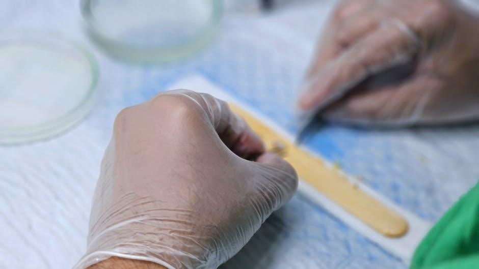
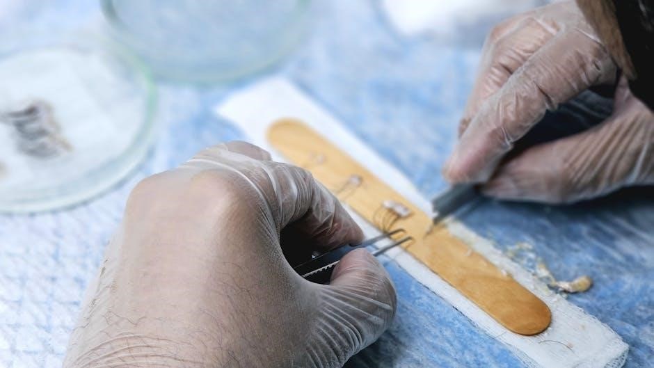
The GTR procedure begins with meticulous scaling and root planing to eliminate biofilm and create a clean root surface. Next, a surgical flap is elevated to access the periodontal defect. The defect is then debrided, removing any remaining inflamed or infected tissue. A resorbable or non-resorbable membrane is carefully positioned over the defect, creating a protected space.
This membrane prevents gingival tissue from entering the defect, allowing bone and periodontal ligament cells to repopulate the area. The membrane is secured with sutures, and the flap is repositioned and sutured closed. Post-operatively, strict oral hygiene is crucial, and regular follow-up appointments are necessary to monitor healing and assess regeneration success. Unlike bone grafting, GTR focuses on soft tissue and attachment apparatus regeneration.
Bone Grafting
Bone grafting, a gold standard for tooth implantation and repair, utilizes autografts, allografts, or xenografts to restore lost bone volume and support dental structures.
Types of Bone Grafts
Bone grafting employs diverse materials categorized into autografts, allografts, and xenografts, each presenting unique characteristics and applications in restorative dentistry. Autografts, considered the gold standard, utilize the patient’s own bone – typically harvested from the hip, tibia, or chin – offering optimal biocompatibility and osteogenic potential, though requiring a secondary surgical site and potential morbidity.
Allografts, sourced from human donors, eliminate the need for a second surgical site but carry a minimal risk of disease transmission, mitigated through rigorous screening and processing. Xenografts, derived from animal sources – commonly bovine – provide a cost-effective alternative, though their osteogenic potential is lower than autografts and allografts, often requiring augmentation with bone morphogenetic proteins or platelet-rich plasma to enhance bone formation. The selection of the appropriate graft type depends on the defect size, patient health, and desired clinical outcome.
Autografts
Autografts represent the “gold standard” in bone regeneration, utilizing bone harvested directly from the patient, typically from intraoral sites like the chin or ramus, or extraoral locations such as the hip or tibia. This approach boasts exceptional osteogenic, osteoinductive, and osteoconductive properties, ensuring robust bone formation and integration due to the patient’s own living cells.
However, autografting isn’t without drawbacks. Harvesting bone necessitates a secondary surgical site, potentially causing donor site morbidity, including pain, swelling, and nerve damage. The limited volume of available bone can also be a constraint for larger defects. Despite these considerations, autografts remain a highly predictable and successful option when sufficient bone volume is available and the patient is a suitable candidate, prioritizing biological compatibility and long-term stability.
Allografts
Allografts involve utilizing bone tissue sourced from a human donor, typically processed by bone banks to ensure safety and minimize immunogenicity. These grafts offer several advantages over autografts, primarily eliminating the need for a second surgical site and providing a potentially larger volume of bone material for reconstruction. Allografts possess osteoconductive properties, providing a scaffold for new bone growth, but generally lack the osteogenic potential of autografts.
While allografts avoid donor site morbidity, concerns regarding disease transmission and immune rejection exist, though modern processing techniques significantly mitigate these risks. They are often combined with other regenerative materials, like growth factors or bone morphogenetic proteins (BMPs), to enhance osteoinductivity and promote faster, more complete bone healing. Careful patient selection and adherence to strict processing protocols are crucial for successful allograft outcomes.
Xenografts
Xenografts utilize bone material derived from non-human sources, most commonly bovine (cow) bone. These materials are extensively processed to remove cellular components and antigens, reducing the risk of immune reactions and disease transmission. Xenografts are primarily osteoconductive, meaning they provide a structural framework for new bone formation, but lack inherent bone-forming cells. This makes them a cost-effective alternative to autografts and allografts, particularly in situations where large bone volumes are not required.
The porous structure of bovine bone promotes vascularization and cellular infiltration, facilitating bone regeneration. Often, xenografts are combined with autologous bone or bone substitutes to enhance their osteogenic potential; While generally safe, potential concerns regarding prion diseases, though extremely rare with current processing methods, exist. Xenografts represent a valuable tool in regenerative dentistry, offering a biocompatible and readily available bone substitute.
Indications for Bone Grafting
Bone grafting becomes essential when significant bone loss compromises dental implant placement or restorative procedures. Common indications include alveolar ridge resorption following tooth extraction, particularly in the posterior regions, and defects caused by periodontal disease or trauma. Patients requiring implant-supported prostheses often necessitate bone augmentation to achieve adequate bone volume and density for successful osseointegration.
Furthermore, bone grafting addresses localized defects such as sinus lifts to create space for implants in the upper jaw, and ridge splits to widen narrow ridges; Correcting bone defects improves aesthetics, function, and long-term implant stability. The need for grafting is determined through clinical examination and radiographic assessment, guiding treatment planning for optimal restorative outcomes. Approximately 2.2 million bone-grafting procedures are performed globally each year.
Bone Grafting Procedure: A Step-by-Step Guide
Step 1: Site Preparation involves meticulous debridement and cleaning of the defect area, ensuring removal of any infected or non-viable tissue. Step 2: Graft Harvesting/Selection determines the source – autograft, allograft, or xenograft – based on defect size and patient needs. Step 3: Graft Placement carefully positions the graft material within the defect, ensuring intimate contact with the existing bone.
Step 4: Membrane Coverage often utilizes a resorbable or non-resorbable membrane to protect the graft and promote bone regeneration. Step 5: Suturing securely closes the surgical site, stabilizing the graft and membrane. Step 6: Post-operative Care includes antibiotics, pain management, and regular follow-up appointments to monitor healing and graft integration. Successful bone grafting relies on precise surgical technique and diligent post-operative care.

GTR vs. Bone Grafting: A Comparative Analysis
Autografting remains the gold standard, yet GTR and GBR, utilizing dental membranes, offer viable alternatives for addressing tooth loss and ridge defects.
When to Choose GTR
Guided Tissue Regeneration (GTR) is particularly indicated when the primary concern is recreating lost periodontal tissues – the gums and supporting structures around the teeth – rather than substantial bone volume. This technique excels in scenarios involving narrow, isolated bony defects, such as those frequently seen after periodontal disease or tooth extraction where sufficient bone height remains.
GTR’s strength lies in preventing soft tissue from encroaching into the bony defect, allowing bone cells to repopulate the area and re-establish a healthy attachment. It’s a preferred approach when the goal is to improve clinical attachment levels and reduce pocket depths. Utilizing dental membranes as a barrier, GTR focuses on soft and hard tissue regeneration in situations where the existing bone foundation is adequate, but soft tissue support is compromised. Therefore, GTR is often selected for furcation defects or intraosseous defects that don’t require significant bone augmentation.
When to Choose Bone Grafting
Bone grafting becomes the treatment of choice when significant bone loss has occurred, creating a volume deficiency that prevents predictable implant placement or restorative procedures. With roughly 2.2 million procedures performed globally each year, it addresses substantial ridge defects resulting from trauma, periodontal disease, or tooth extraction. The ‘gold standard’ – autografting – utilizes the patient’s own bone, ensuring biocompatibility and optimal integration.
If a large bony defect exists, or if the goal is to increase ridge width or height to support a dental implant, bone grafting is the preferred solution. It provides a foundational scaffold for new bone formation, creating a stable base for future dental work. Unlike GTR, which focuses on guided tissue regeneration within an existing bone structure, bone grafting aims to reconstruct missing bone volume, making it essential for cases with severe bone resorption.
Combining GTR and Bone Grafting
A synergistic approach often involves combining GTR and bone grafting, particularly in cases with both significant bone loss and compromised soft tissue. Initially, bone grafting reconstructs the lost alveolar ridge volume, establishing a solid foundation. Subsequently, GTR techniques are employed to prevent soft tissue ingrowth into the newly grafted bone, ensuring the preservation of the desired space for periodontal ligament regeneration and ultimately, implant stability.
This sequential or simultaneous approach maximizes regenerative potential. Bone grafts provide the bulk, while GTR membranes act as a barrier, guiding the healing process. This is especially beneficial in complex defects where both hard and soft tissue regeneration are critical for long-term success. The combined strategy leverages the strengths of each technique, offering a more predictable outcome than either procedure alone, particularly when addressing extensive ridge defects.
Recent Advancements in Regenerative Dentistry
3D printing revolutionizes GTR and bone grafting, creating custom structures, while growth factors enhance regeneration, shaping the future of ridge reconstruction.
3D Printing in GTR and Bone Grafting
Over the past decade, 3D printing has transitioned from a futuristic concept to a revolutionary tool within medicine, particularly impacting regenerative dentistry. Its remarkable ability to produce custom-made, complex structures is fundamentally changing how clinicians approach treatments like Guided Tissue Regeneration (GTR) and Bone Grafting.
Traditionally, GTR relied on standardized membranes. However, 3D printing allows for the creation of patient-specific membranes precisely tailored to the defect’s morphology, optimizing tissue integration and regeneration. Similarly, in bone grafting, 3D-printed scaffolds can be designed with specific porosity and architecture to encourage bone ingrowth and vascularization. This personalized approach enhances the predictability and success rates of these procedures.
Furthermore, 3D printing facilitates the incorporation of growth factors directly into the scaffolds, providing a localized and sustained release to further stimulate tissue repair. This technology promises to minimize surgical time, improve aesthetic outcomes, and ultimately, offer more effective solutions for patients experiencing tooth loss and ridge defects.
The Role of Growth Factors
Growth factors are increasingly recognized as pivotal components in enhancing the efficacy of both Guided Tissue Regeneration (GTR) and Bone Grafting procedures. These biologically active molecules stimulate cellular proliferation, differentiation, and angiogenesis – crucial processes for successful tissue regeneration. In GTR, growth factors like Platelet-Derived Growth Factor (PDGF) can promote periodontal ligament cell migration and attachment, accelerating the formation of a new periodontal apparatus.
Similarly, in bone grafting, Bone Morphogenetic Proteins (BMPs) are frequently utilized to induce osteoblast differentiation and new bone formation. The localized and sustained release of these factors, often achieved through incorporating them into scaffolds or membranes, optimizes their therapeutic effect.
Recent advancements focus on combining multiple growth factors to mimic the natural healing cascade, leading to more comprehensive and predictable regenerative outcomes. This approach represents a significant step towards personalized regenerative dentistry.
Future Trends in Ridge Regeneration
Future trends in ridge regeneration are heavily focused on personalized approaches, leveraging advancements in 3D printing and biomaterials. Expect to see more patient-specific scaffolds designed to precisely fit defects, incorporating tailored combinations of growth factors for optimized healing. Research is expanding beyond traditional GTR and bone grafting, exploring the potential of stem cell therapies and gene editing to enhance regenerative capacity.
Furthermore, minimally invasive techniques and digital workflows will become increasingly prevalent, reducing patient morbidity and improving predictability. Smart biomaterials capable of responding to the biological environment, releasing therapeutic agents on demand, are also on the horizon.
Ultimately, the goal is to create regenerative procedures that are less reliant on donor sites and yield more natural, functional, and aesthetically pleasing results.